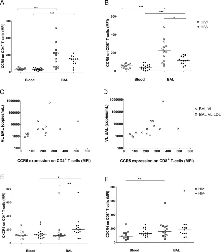
Figure 2.

CCR5 and CXCR4 expression on CD4+ or CD8+ T cells in blood and BAL. (A, B, E, F) The median fluorescence intensity (MFI) of (A, B) CCR5 and (E, F) CXCR4 receptor expression was measured by flow cytometry on (A, E) CD4+ and (B, F) CD8+ cells from the blood and BAL of HIV-1-infected (n = 12 in blood, n = 14 in BAL, open squares) and HIV-1-uninfected persons (n = 16 in blood, n = 14 in BAL, solid circles), each sign represents one individual, bars represent medians. Differences between CCR5 expression on CD4+ and CD8+ T cells from paired blood and BAL samples were calculated by Wilcoxon signed rank test (***p < 0.001). The CCR5 expression on CD8+ T cells in the BAL was significantly higher in HIV-1-infected compared to HIV-1-uninfected persons (*p = 0.026, by Mann–Whitney U-test). (C, D) The correlation between viral load (VL) in BAL and MFI of (C) CCR5+ on CD4+ (ρ = 0.706, p = 0.005) or (D) CD8+ (ρ = 0.793, p < 0.001) BAL T cells of HIV-1-infected participants was assessed by Spearman. Open circles, BAL VL; symbol ×, BAL VL LDL were set to a value of 19 copies/mL and normalised according to the urea method. (E) The difference of CXCR4 expression on CD4+ paired blood and BAL T cells was measured by Wilcoxon signed rank test (*p = 0.049). MFI of CXCR4+ CD4+ BALMCs was significantly higher in the HIV-1-uninfected control group when compared with that of HIV-1-infected persons (**p = 0.009, Mann–Whitney U-test). (F) BAL CD8+ T cells expressed higher levels of CXCR4 than PBMCs in HIV-1-infected persons (**p = 0.003), differences between the HIV-1 status were assessed by Mann–Whitney U-test. Data shown are pooled data from experiments on 12 blood and 14 BAL samples of HIV-1-infected and 16 blood and 14 BAL samples of HIV-1-uninfected persons.
