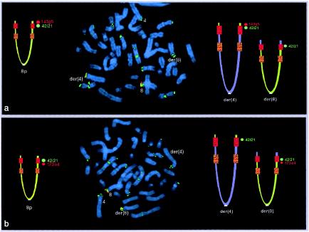
Figure 3.
Metaphase FISH in case subject 7 (with the 46,XY,t(4;8)(p16;p23.1) translocation), showing the der(8) breakpoint. In the ideograms, the normal 8p is on the left, and the two derivative chromosomes are on the right; the red and orange squares indicate the REPD and the REPP, respectively. Green signals correspond to GS-42i21, which maps within the 8p REPD; these signals are present in several OR-gene cluster regions. a, Probe GS-143g5 (red signals), which is distal but adjacent to the 8p REPD. This clone is transposed to the der(4). b, Probe GS-173o4 (red signals), which is the most distal clone within the region included between the two 8p REPs. This clone remains on the der(8). These data demonstrate that the 8p breakpoint is within the REPD (see also fig. 1 in Eichler 2001).

