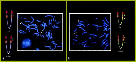
Figure 4.
FISH in the mother of case subject 3 (with 46,XX). a, Normal and inverted 4p. At left, the ideograms show that clones RP11-323f5 (red signals) and RP11-448g15 (green signals), included between the two 4p REPs, are inverted in the chromosome 4 (arrowhead). In the square, a nucleus shows the same inversion; yellow signals refer to the control probe RP11-520m5, which is distal to the 4p REPD (see fig. 1), and red and green signals refer to RP11-323f5 and RP11-190l6, respectively (these two clones are at a distance of ∼2 Mb and thus are more appropriate for interphase FISH). b, Normal and inverted 8p. At right, the ideograms show that clones GS-173o4 (red signals) and GS-257o3 (green signals), included between the two 8p REPs, are inverted in one chromosome 8 (arrowhead). RP11-563o19 (yellow signals), at 8p12, was used as a control probe.

