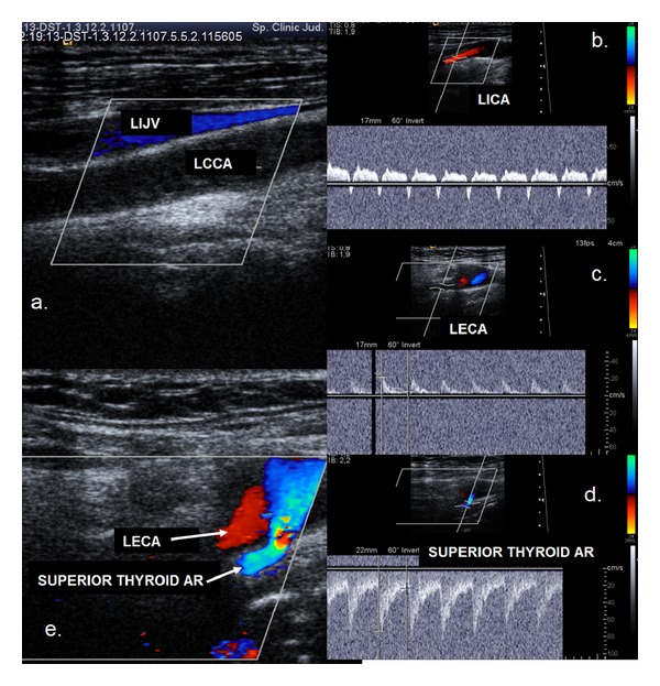
Figure 2.

Ultrasound examination showing common carotid artery occlusion with patent distal vessels. Anterograde flow in both the ICA and ECA. (a) Colour mode examination: no flow in the left common carotid artery (LCCA), and the vessel lumen is filled with thrombotic material. (b) Duplex mode examination: anterograde flow in the left internal carotid artery with a steal effect, deceleration, and inversed flow in mesosystole. (c) Duplex mode examination: anterograde flow in the left external carotid artery. (d) Duplex mode examination: retrograde flow in the left superior thyroid artery. (e) Colour mode examination: retrograde flow in the left superior thyroid artery and anterograde flow in LECA.
