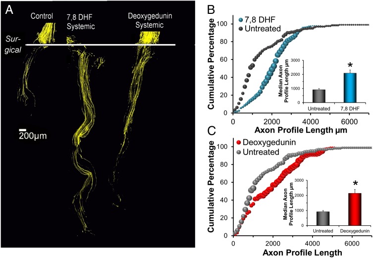
Fig. 4.
(A) Branches of the sciatic nerve of thy-1-YFP-H mice were cut and repaired with grafts of the same nerve obtained from WT, non-YFP-expressing littermates and were treated daily by i.p. injections of either 7,8 DHF or deoxygedunin or were untreated (Control). Each image is a compilation of stitched images from single 10-μm-thick optical sections through the graft. The proximal cut nerve segments are aligned at the top of each panel. The horizontal line marks the site of surgical repair. (B and C) Distributions of axon profile lengths in these mice. Data from systemically treated mice are compared with similar data from untreated mice, the same data as shown in Figs. 1–3. (Insets) Average median axon profile lengths (+SEM).

