Fig. 2.
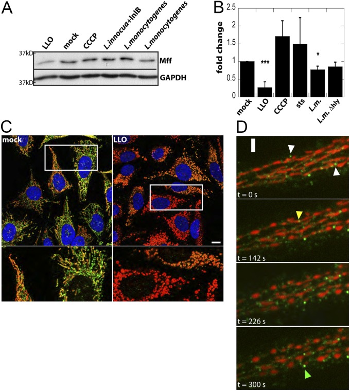
LLO treatment and L. monocytogenes infection cause a decrease in the Drp1 receptor Mff. (A) HeLa cells were treated for 10 min with 6 nM LLO, 30 min with 10 µM CCCP, or infected at an MOI of 50 with the indicated strains for 1 h. Representative Western blot of Mff and GAPDH (loading control) on total cell lysates shows a decrease in Mff levels upon LLO treatment or infection with wild-type L. monocytogenes. (B) Quantification of Western blots from >3 independent experiments (***P < 0.001; *P < 0.05). (C) Immunofluorescence of HeLa cells treated for 10 min with LLO and stained for Mff (green), mitochondria (Tom20; red), and DNA (DAPI; blue). Although there is heterogeneity in staining of endogenous Mff, there is a significant decrease in Mff staining upon LLO treatment. White box indicates a 4× enlarged region that is shown below. (D) Heterogeneous behavior of mitochondria-associated Mff puncta observed by live cell imaging. Timelapse spinning disk confocal images of HeLa cells expressing Mff-GFP (green) and TagBFP-mito (red). Data were collected every 2 s 2 nM LLO was added at time t = 50 s White arrowheads mark Mff-GFP puncta that are rapidly lost from mitochondria. Yellow arrowhead marks an Mff dot that disappears before fragmentation and green arrowhead marks one that remains associated with mitochondria over the time course. (Scale bars: C, 10 μm; D, 2.6 μm.)

