Fig. 3.
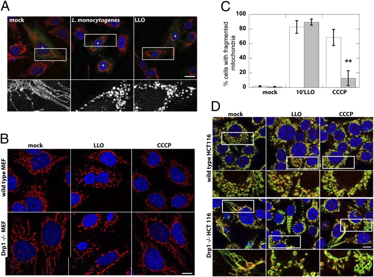
LLO and L. monocytogenes induce mitochondrial-fragmentation in Drp1−/− cells. (A) Infection of HeLa cells transfected with K38A-HA Drp1 with L. monocytogenes for 1 h or treatment with 6 nM LLO for 10 min induces mitochondrial fragmentation. Immunofluorescence images show mitochondria (CoxIV; red) and K38A-HA Drp1 (anti-HA; green). Asterisk marks transfected cells, as identified by immufluorescence. The white box indicates the portion of the image enlarged 4× and shown below. (Scale bars: 20 μm.) (B) Immunofluorescence of mitochondria (CoxIV; red) and nuclei (DAPI; blue) upon treatment of WT or Drp1−/− MEFs with either 6 nM LLO (10 min) or 10 µM CCCP (30 min). (C) The percentage of cells displaying a fragmented mitochondrial network upon CCCP or LLO treatment was quantified in five or six randomly chosen fields of view (n > 150 cells) per experiment. Pooled data from three independent experiments is shown (**P < 0.003, one-tailed Student t test). (D) Immunofluorescence of mitochondrial matrix (cytochrome c; red), mitochondrial outer membrane (Tom20; green), and nuclei (DAPI; blue) upon treatment of WT or Drp1−/− HCT116 cells with either 6 nM LLO (10 min) or 10 µM CCCP (30 min). White box indicates 2× enlarged region shown below. (Scale bars: 10 μm.)

