Fig. 1.
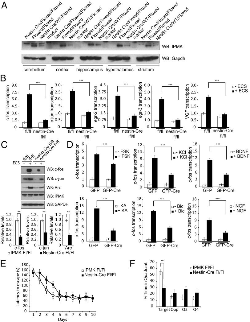
IPMK enhances IEG expression. (A) Nestin-Cre IPMKfl/fl mice display uniform depletion of IPMK in cerebellum, cortex, hippocampus, hypothalamus, and striatum of the brain. Nestin-Cre IPMKwt/fl mice exhibit half the levels of IPMK present in each brain region tested, reflecting deletion of only the floxed allele. (B) Levels of IEG expression as assessed via quantitative PCR (qPCR) are increased in the hippocampus of IPMKfl/fl mice after ECS. In nestin-Cre IPMKfl/fl mice, levels of IEG expression are decreased 60%–80%. ***P < 0.001. Data are means ± SEM from four experiments. Data are expressed as fold over baseline, where baseline is defined as IPMKfl/fl mice without ECS treatment. (C) Western blotting of hippocampal tissue in IPMKfl/fl mice after ECS reveals robust enhancement of c-fos, c-jun, and Arc protein levels. In nestin-Cre IPMKfl/fl mice, levels of IEG expression are decreased 50–70%. **P < 0.01. Data are means ± SEM from four experiments. (D) Primary cortical neurons from IPMKfl/fl mice are infected with lentivirus expressing GFP or GFP-Cre and treated with KCl, forskolin (Fsk), NGF, BDNF, bicuculline (Bic), and kainic acid (KA). In all circumstances, neurons with IPMK deletion exhibit impaired c-fos mRNA induction. ***P < 0.001, Student t test. Data are means ± SEM from three experiments. (E) Performance on the Barnes maze task during training for 10 d (two trials per day, 1 h intertraining interval). Nestin-Cre IPMKfl/fl (n = 16) mice exhibit higher latencies to find the exit versus control littermates (n = 18) during the initial phase of training. F1,32 = 4.24, P < 0.05; F1,32 = 5.37, P < 0.05; F1,32 = 7.80, P < 0.01. (F) Nestin-Cre IPMKfl/fl mice show a deficiency in spatial memory during a probe trial performed 1 wk after training. IPMK brain knockout mice are significantly impaired in spatial localization for the correct quadrant. F1,32 = 5.88, P < 0.05.

