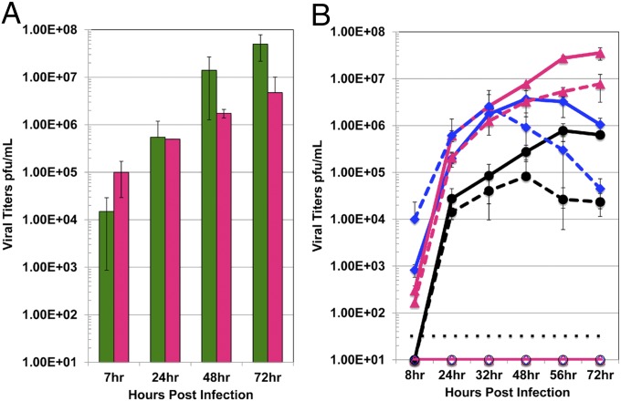
Fig. 5.
Recombinant virus growth in primary human lung cells. HAEs, primary alveolar type II pneumocytes, primary lung microvascular endothelial cells, and primary lung fibroblast cells were infected with wild-type rSARS-CoV, wild-type MERS-CoV, or rMERS-CoV-RFP. (A) Wild-type MERS-CoV and SARS-CoV growth kinetics in HAEs. Data are representative of two independent experiments. Error bars represent SD from the mean. Green bars, wild-type SARS-CoV; pink bars, wild-type MERS-CoV. (B) Wild-type SARS-CoV, wild-type MERS-CoV, and rMERS-CoV-RFP growth kinetics were compared in alveolar type II (ATII) pneumocytes, lung microvascular endothelial (MVE) cells, and lung fibroblasts (FBs). SARS-CoV did not replicate in any of the primary cell types examined in B. Cells were specific to one of two donors, and neither was permissive for SARS-CoV. Data are representative of two independent experiments. Error bars represent SD from the mean. The dotted black line in B represents the limit of detection for the assay. All points under this line are considered to be negative for replication in the samples assayed. Solid blue line with closed diamond, wild-type MERS-CoV in ATII cells; dotted blue line with closed diamond, rMERS-CoV-RFP in ATII cells; solid blue line with open diamond, wild-type SARS-CoV in ATII cells (did not replicate); solid pink line with closed triangle, wild-type MERS-CoV in FB cells; dotted pink line with closed triangle, rMERS-CoV-RFP in FB cells; solid pink line with open triangle, wild-type SARS-CoV in FB cells (did not replicate); solid black line with closed circle, wild-type MERS-CoV in MVE cells; dotted black line with closed circle, rMERS-CoV-RFP in MVE cells; solid black line with open circle, wild-type SARS-CoV in MVE cells (did not replicate).

