Fig. 1.
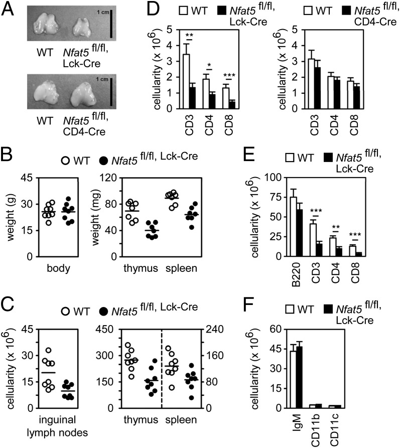
Reduced number of thymocytes and peripheral T cells in Nfat5fl/fl, Lck-Cre mice. (A) Thymus size in control (WT), Nfat5fl/fl, Lck-Cre (Upper), or Nfat5fl/fl, CD4-Cre (Lower) mice. (Scale bar, 1 cm.) (B) Body, thymus, and spleen weight in WT and Nfat5fl/fl, Lck-Cre mice. (C) Cell numbers in inguinal lymph nodes, thymus, and spleen in WT and Nfat5fl/fl, Lck-Cre mice. Each circle in B and C represents a single mouse, and the horizontal lines show the mean. (D) Numbers of CD3, CD4, and CD8 cells in lymph nodes of WT and Nfat5fl/fl, Lck-Cre mice (Left) and WT and Nfat5fl/fl, CD4-Cre mice (Right) (mean ± SEM); n ≥ 6. Numbers of B220-positive, CD3-positive, CD4-positive, and CD8-positive (E) and IgM-positive, CD11b-positive, and CD11c-positive (F) splenocytes in WT and Nfat5fl/fl, Lck-Cre mice (mean ± SEM); n = 8. *P < 0.05; **P < 0.01; ***P < 0.001.

