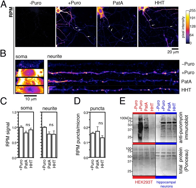
Fig. 3.
Neuronal polyribosomes are stalled downstream of the first round of translation elongation. (A) Treatment with drugs that induce ribosome runoff reveals the persistence of RPM puncta (arrows). (B) Magnified images of soma and straightened neurites from confocal images in A. (C) Quantified RPM signal in soma and neurites from images represented in B. (D) Quantified number of RPM puncta in neurites found more than 50 μm from the nucleus (refer to Materials and Methods for parameters). (E) Comparison of puromycylated peptides between nonneuronal HEK293T cells and primary rat hippocampal neurons by Western blot analysis following RPM (Upper). Total protein levels were assessed by Ponceau staining (Lower). Blot is representative of three independent experiments. All bars represent mean ± SEM and n = 9 cells from three independent experiments.

