
Figure 2.
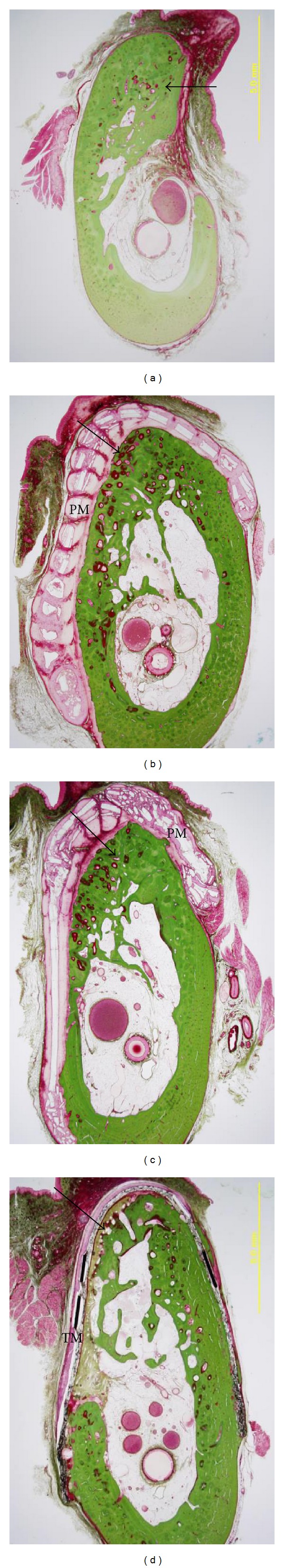
Histological microphotographs of coronal sections at 6 months postoperatively [33]. The Villanueva-Goldner staining: (a) control (virgin) group, (b) GBR using PLGC membrane, (c) GBR using PLGC+bone chips group, and (d) GBR using TR-PTFE membrane. PM: PLGC macroporous membrane. TM: TR-PTFE membrane, and arrow: regenerated bone.