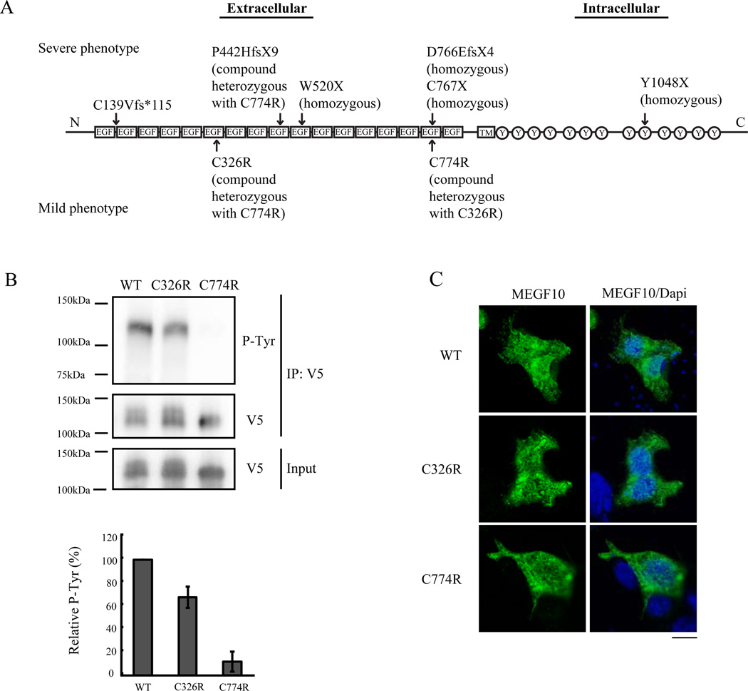
Figure 1. Tyrosine phosphorylation in C326R and C774R mutant MEGF10.
(A) Diagram of the MEGF10 protein domains with arrows indicating previously reported human patients’ mutations [2,3,15]. Upper mutations are from individuals with severe disease phenotypes. Lower mutations are from three patients in one family with a mild phenotype. Note that the C774R mutation has been associated with both severe and mild phenotypes. EGF: EGF-like domain, TM: transmembrane domain, Y: tyrosine residue. (B) HEK293T cells were transfected with wild type, C326R mutant, and C774R mutant MEGF10 tagged with V5. Cell lysates were immunoprecipitated with anti-V5 antibody, then subjected to immunoblotting with anti-phosphotyrosine (P-Tyr) antibody or anti-V5 antibody. The C774R mutant shows a greater defect in tyrosine phosphorylation than the C326R mutant. IP V5: V5 tagged immunoprecipitated lysates. Western blot densitometry of the bands shows decreased tyrosine phosphorylation in C326R and C774R. Quantitative analysis was normalized against intensities of immunoprecipitated V5 bands (data are relative to wild type MEGF10, n=5). (C) Wild type, C326R mutant, and C774R mutant MEGF10 constructs were transfected into 293T cells. One day after transfection, cells were fixed and stained with anti-V5 antibody (green). Representative cells are shown. The left column shows MEGF10 staining with anti-V5 antibody. The right column shows merged images of DAPI-labeled nuclei (blue) with the images on the left. Mutant proteins show the same subcellular localization pattern as the wild type proteins. Scale bar: 10µm.

