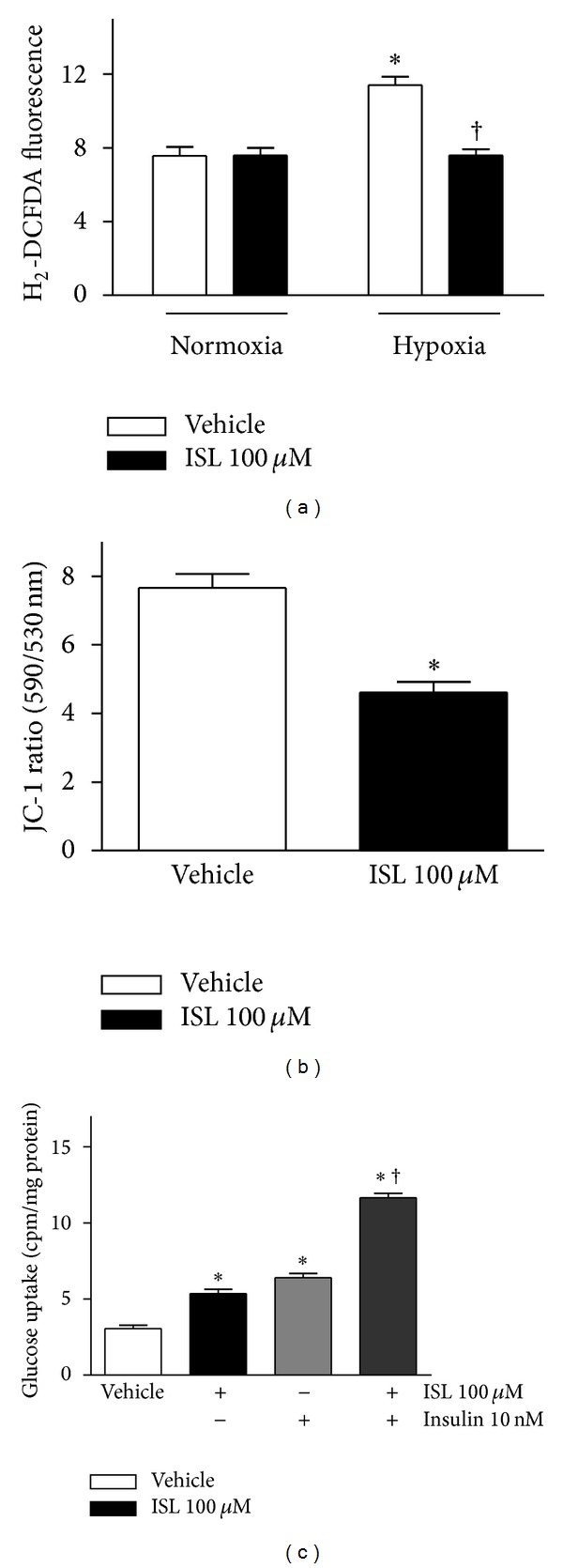
Figure 4.

(a) ISL reduced the intracellular ROS levels in isolated mouse cardiomyocytes during hypoxia/reoxygenation. Intracellular ROS levels were measured by the fluorescent probe H2DCFDA after treatment with ISL (100 μM) or DMSO (vehicle). ROS production was expressed as fluorescence intensity relative to untreated control cells. Data are presented as means ± SE (n = 4–6). *P < 0.05 versus normoxia vehicle; † P < 0.05 versus hypoxia vehicle; (b) ISL reduced mitochondrial membrane potential (Δψ) in isolated cardiomyocytes. Mitochondrial membrane potential (Δψ) was measured by JC-1 fluorescence assay. The result was presented as the ratio of red/green fluorescence measured at 590 nm and 530 nm, respectively. Values are means ± SE (n = 6–10). *P < 0.01 versus vehicle; (c) ISL treatment augmented glucose uptake of cardiomyocytes. The cardiomyocytes were preincubated for 30 min with or without ISL (100 μM) and/or insulin (10 nM), before addition of 2-deoxy-[1-3H]glucose for additional 30 min to measure glucose uptake. Values are means ± SE for 5 experiments. *P < 0.05 versus vehicle; † P < 0.05 versus insulin alone.
