Figure 3.
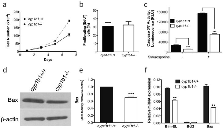
Lack of Cyp1B1 in retinal PC resulted in increased proliferation and decreased apoptosis. (a) The rate of cell proliferation was increased in cyp1b1−/− PC compared to wild-type cells by counting the cell numbers. (b) Cyp1b1+/+ and cyp1b1−/− PC display similar rates of DNA synthesis by flow cytometry (P > 0.05). (c) The rate of apoptosis was determined by measuring caspase activity with luminescent signal from caspase-3/7 DEVD-aminoluciferin substrate. Cyp1b1−/− PC demonstrated a 2 -fold decrease in basal levels of caspase-3/7 and a 2-fold decrease when challenged with 10 nM staurosporine. RLU, relative luminescence unit. (d) Expression of Bax was analyzed by Western blotting. The β-actin level was assessed as a loading control. (e) Quantification of band intensity demonstrated a 1.4-fold decrease in Bax expression in the cyp1b1−/− PC. (**P< 0.001, ***P<0.0001). (f) Relative mRNA expression of Bim-EL, Bax, and Bcl-2 was analyzed by real-time PCR (N=3, **P<0.001)

