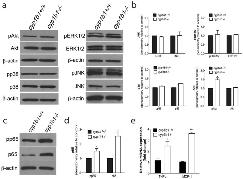
Figure 9.
Alterations in cellular signaling pathways in cyp1b1−/− retinal PC. (a) Cyp1b1+/+ and cyp1b1−/− PC were analyzed by Western blot analysis for expression of phospho -Akt, total Akt, phospho-p38, total p38, phospho-Erk1/2, total Erk1/2, phospho-JNK, total JNK and β-actin. (b) Quantification of band intensity demonstrated a 1.5 fold increase in phospho-Akt (N=3, *P < 0.05). (c) Levels of phospho-p65 NF-κB, total p65, and β-actin were determined by Western blotting. (d) Quantitative assessment of the data (N=3, *P<0.05). (d, e) Levels of RNA were assessed for NF-κB target genes MCP-1 (***P ≤ 0.0001) and TNFα (*P<0.05).

