Figure 5.

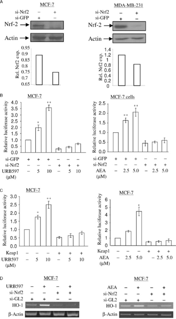
Depletion of Nrf2 abolishes URB597- and AEA-induced nqo1-ARE-Luc reporter activity and HO-1 mRNA expression. (A) Depletion of Nrf2 protein by siRNA approach in MCF-7 cells and MDA-MB-231 cells. MCF-7 cells and MDA-MB-231 cells were treated with 5 nM of si-GFP RNA (control) and si-Nrf2 RNA as indicated. At 48 h, 15 μg of total cell lysates was subjected to immunoblotting in order to detect the levels of Nrf2 protein. The same membrane was blotted with anti-actin antibody to monitor equal loading of proteins. The levels of Nrf2 protein were normalized to actin levels and the relative expression was represented graphically. All the figures are representative of three independent experiments. (B) Depletion of Nrf2 blocks URB597- and AEA-induced nqo1-ARE-Luc reporter activity. MCF-7 cells were treated with 5 nM of si-GFP RNA (control) and si-Nrf2 RNA as indicated. At 16 h, the cells were introduced with 0.5 μg of nqo1-ARE-Luc reporter and 0.15 μg of pCMV-β-galactosidase plasmids, followed by URB597 (5.0 μM) and AEA (2.5 μM) treatment for 16 h. Luciferase activity assay and data analysis were performed as in Figure 1C. The data represent the mean ± SD of at least three independent experiments performed in triplicate. *P < 0.05 as compared with the cells treated with siGFP; **P < 0.001 as compared with cells treated with si-GFP. (C) Keap1 blocks URB597- and AEA-induced nqo1-ARE-Luc reporter activity. Furthermore, 0.5 μg of nqo1-ARE-Luc reporter and 0.15 μg of pCMV-β-galactosidase plasmid were introduced into MCF-7 cells with or without Keap1, followed by 5.0 μM of URB597 treatment and 2.5 μM of AEA treatment. After 16 h, luciferase activity assay and data analysis were performed as in Figure 1C. The data represent the mean ± SD of at least three independent experiments performed in triplicate. *P < 0.05 as compared with the untreated cells; **P < 0.001 as compared with untreated cells. D. MCF-7 cells were treated with 5 nM of si-GL2 RNA (control) and si-Nrf2 RNA as indicated. At 48 h, cells were treated with 5 μM of URB597 or 2.5 μM of AEA for 6 h as indicated. Semi-quantitative RT-PCR for HO-1 mRNA was performed as in Figure 1A. All figures are representative of three independent experiments.
