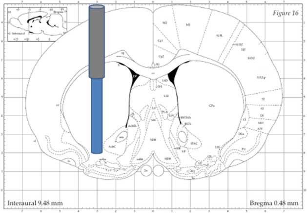
Figure 1.

Illustration ofmicrodialysis probe placement in dorsal and ventral striatum. The guide cannula was placed through a hole in the skull at the following coordinates relative to bregma: anterior + 0.5 mm, lateral ± 3.0 mm, and lowered 4.0 mm ventral relative to bregma, microdialysis probe protruding additional 4.0 mm ventral relative to bregma (Paxinos and Watson, 1998).
