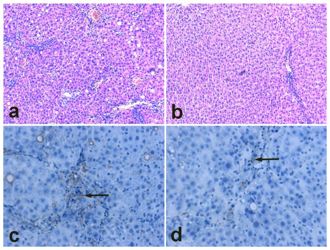
Figure 9. Micrographs of histological specimens of liver tissue (×200).
Liver fibrosis was successfully induced by CCL4 after 10 weeks. Hematoxylin-eosin staining shows fibrotic (a) and normal (b) rat livers. Compared with the nontargeting group (d), more EGFP-positive cells were observed in the targeting group (c).

