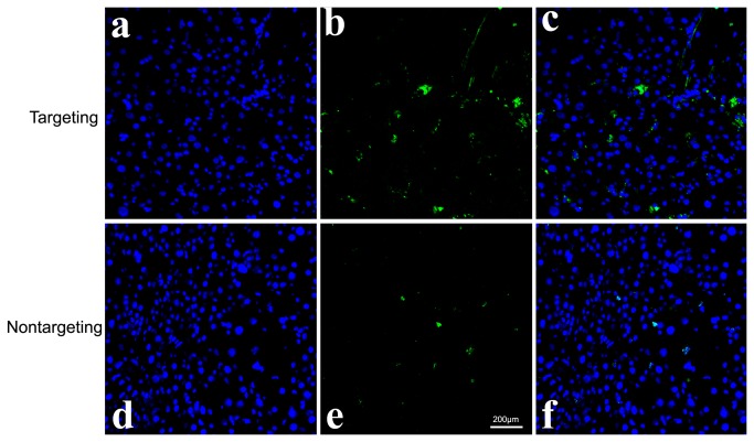
Figure 10. Confocal laser scanning microscopic images of frozen sections.
a, b, c) targeting group; d, e,f) nontargeting group. Images assignment: green: EGFP fluorescence, blue: nucleus. The image on the right is an overlay of the two fluorescent colors. Confocal laser scanning microscopic images of frozen sections showed that more EGFP fluorescence was observed in the targeting group.

