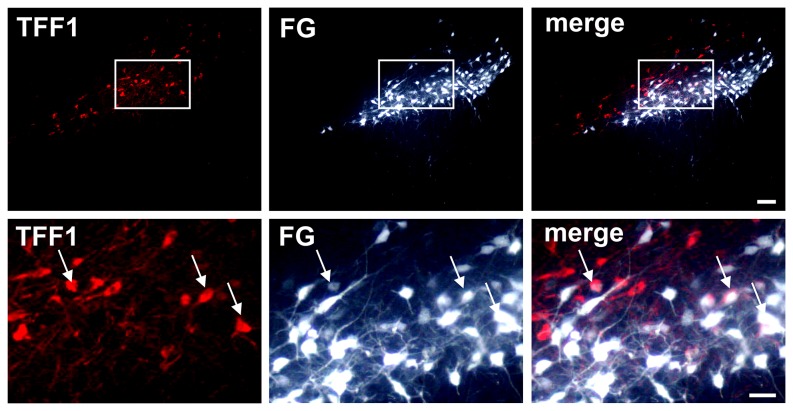
Figure 7. Detection of Trefoil factor 1 (TFF1) in nigrostriatal projection neurons by Fluorogold (FG) labelling.
Representative photomicrographs of double immunofluorescence stainings for TFF1 and the retrograde tracer FG at the level of substantia nigra pars compacta (SNc) 10 days after intrastriatal FG injection. A subpopulation of TFF1-ir cells was found to co-express FG identifying these as projection neurons (arrows). Scale bars upper panel: 100 µm, lower panel: 50 µm.

