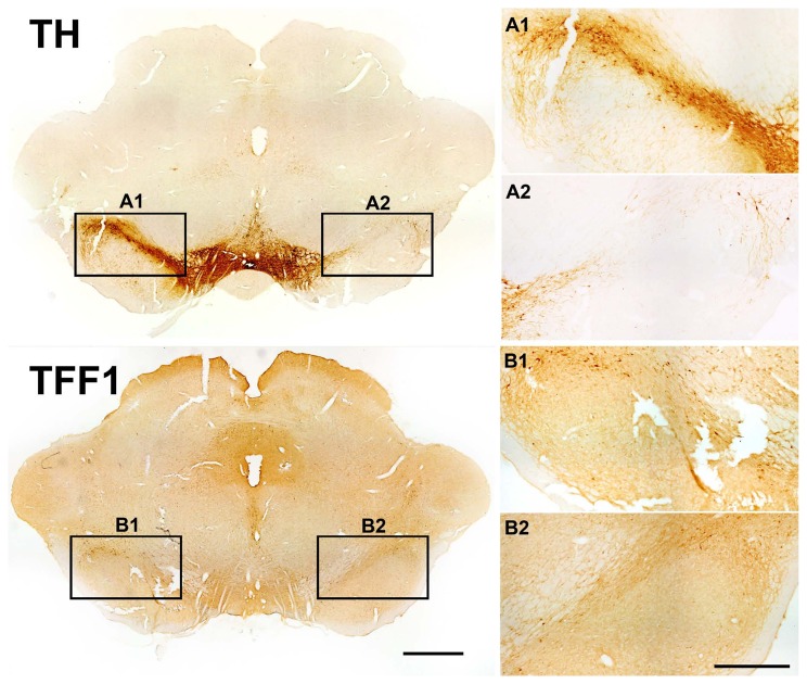
Figure 8. Loss of trefoil factor 1 (TFF1) expressing cells in a rat model of Parkinson’s disease.
Representative photomicrographs of tyrosine hydroxylase (TH; A) and TFF1 (B) in brain sections from adult rat ventral mesencephalon at 4 weeks after unilateral 6-hydroxydopamine (6-OHDA) lesion. The lesion resulted in a distinct loss of TH-ir neurons in right SN (A2) as compared to the contralateral, unlesioned control side (A1). Similarly, a reduction of TFF1-ir cells was detected on the lesioned side (B2) as compared to the intact control side (B1). This loss of TFF1-ir cells is better recognized on the enlarged images (B2). Scale bars: 1 mm (overview), 500 µm (magnification).

