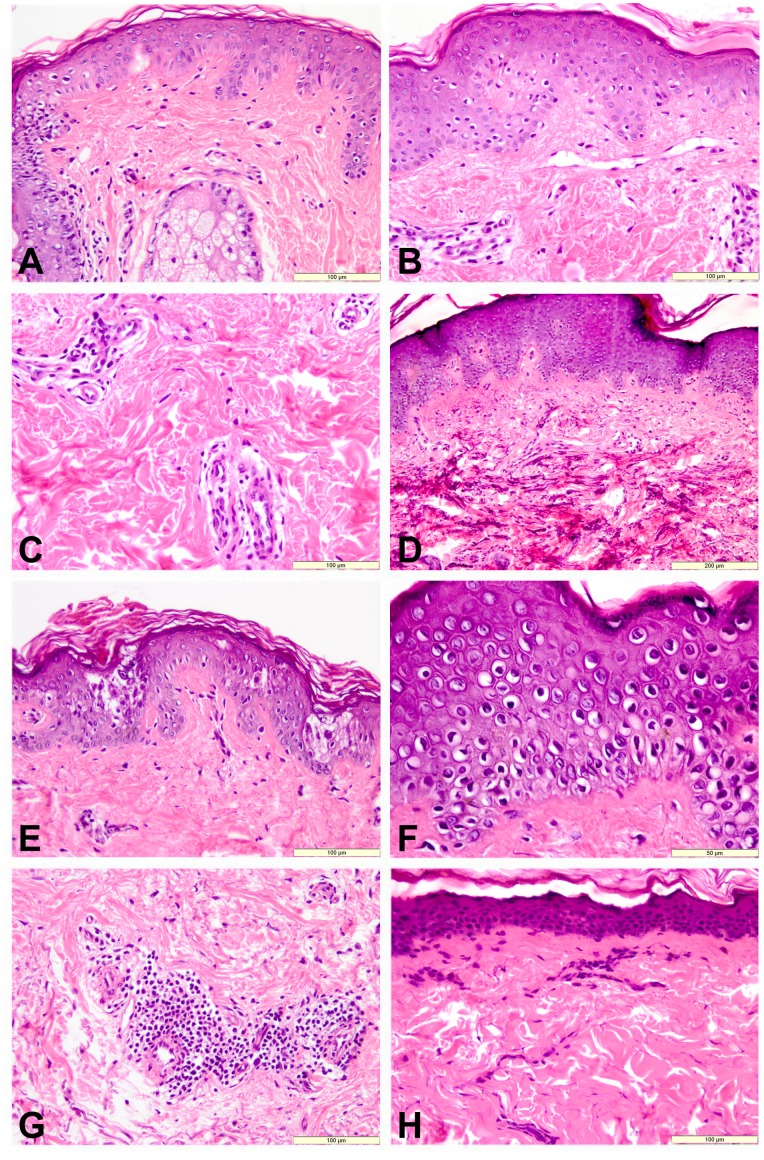
Figure 1. Histological aspects of the skin from different groups and subgroups.
A. Group 1, UV non-protected regions 1. Intracellular edema and scattered inflammatory cells. HE, bar = 100 µm; B and C. Group 2, UV non-protected regions; intracellular edema, perivascular edema and mononuclear inflammatory cells, dermal fibrosis. HE, bar = 100 µm; D and E. Group 3 , UV non-protected regions. Intracellular edema, dermal fibrosis and mineralization of collagen fibers; HE, bar = 200 µm; multiple foci of epidermal necrosis. HE, bar = 100 µm; F and G. Group 4, UV non-protected regions. F - Diffuse intracellular edema of epidermis and multiple apoptotic cells. HE, bar = 100 µm; G - Dermal fibrosis and perivascular mononuclear cell aggregation. HE, bar = 100 µm; H. Group 4 , UV protected regions. Hyperkeratosis, epidermal atrophy and uniform dermal fibrosis. HE, bar = 100 µm.

