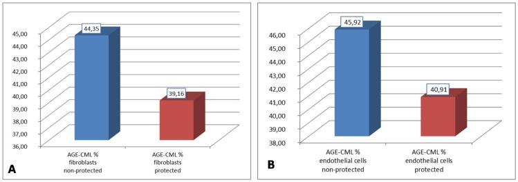
Figure 3. AGE-CML expression in fibroblasts and endothelial cells from both UV protected and non-protected sites.
(A) AGE-CML is statistically higher in non-protected fibroblasts than in protected tissues (paired t-test probability is less than 0.001); (B) AGE-CML is statistically higher in non-protected endothelial cells than in protected tissues (paired t-test probability is less than 0.001).

