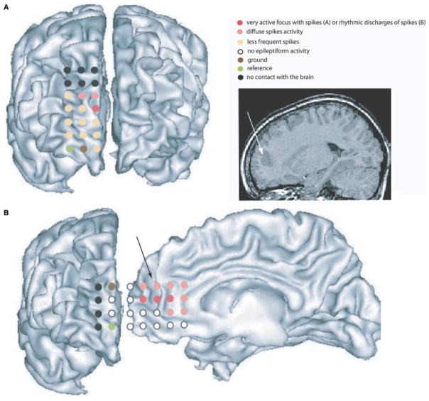
Fig. 2.
Electrocorticographic recording for Patient 5. The white arrow on theT1 MRI and the black arrow on the reconstructed cortical surface show the dysplastic sulcus in the right anterior cingulate gyrus. (A) A 3 × 8 grid was positioned over the right frontopolar area. The two upper rows of electrodes did not touch the brain (contacts in black). A spiking activity was clearly observed on the red contact, more diffuse over the contacts in pink. Rare spikes were noticed over the orange contacts. Pink and red contacts were in front or in close vicinity of the dysplastic sulcus. (B) A 4 × 8 grid was inserted in the interhemispheric space in contact with the right anterior cingulate area. The last two rows of electrodes were curved and only one was in contact with the frontopolar area. Contacts in red showed prolonged rhythmic discharges of spike activity and were in front of the bottom of the dysplastic sulcus. In pink, contacts showed diffuse spiking activity.

