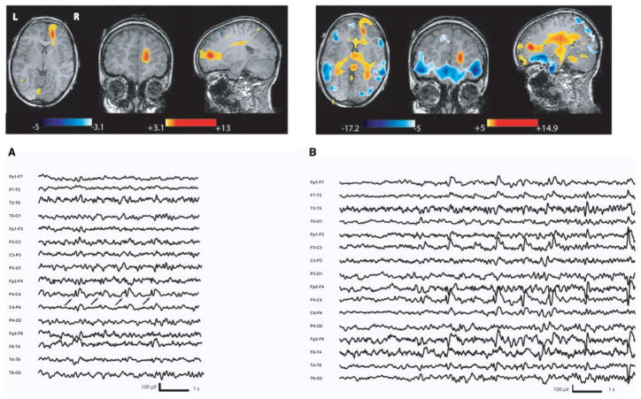
Fig. 5.
Focal cortical dysplasia (Patient 5). Figure 2 showed in the anatomical MRI a right dysplastic sulcus in the anterior cingulate gyrus (white arrow). (A) Activation in the dysplasia during interictal right frontal spike and waves. (B) BOLD signal changes observed during clinical seizures with prolonged rhythmic discharge of right frontal spike and waves with synchronous contralateral activity. Maximum activation is again in the lesion with propagation over the right frontopolar area, thalami and the midbrain (left frontopolar area, left anterior cingulate area were also involved but not shown on this picture).

