Fig. 7.
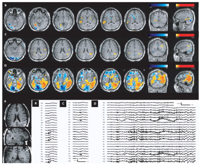
Band heterotopia (Patient 7). (A) Anatomical MRI showed a bilateral posterior band heterotopia (head of white arrows). (B) Activation in the left band heterotopia and overlying cortex duringT5–P9– O1polyspikes. (C) Activation of the right band heterotopia duringT6–P10– O2 polyspikes (deactivation, blue, in both occipital lobes). (D) Maximum activation during seizures seen in right and left band heterotopia and overlying cortex (deactivation in blue). Ictal EEG onset showed right posterior temporal fast activity followed by left posterior temporal fast activity. In interictal and ictal events, heterotopic and normotopic cortices are involved by BOLD changes. These BOLD changes are predominant in the heterotopic cortex.

