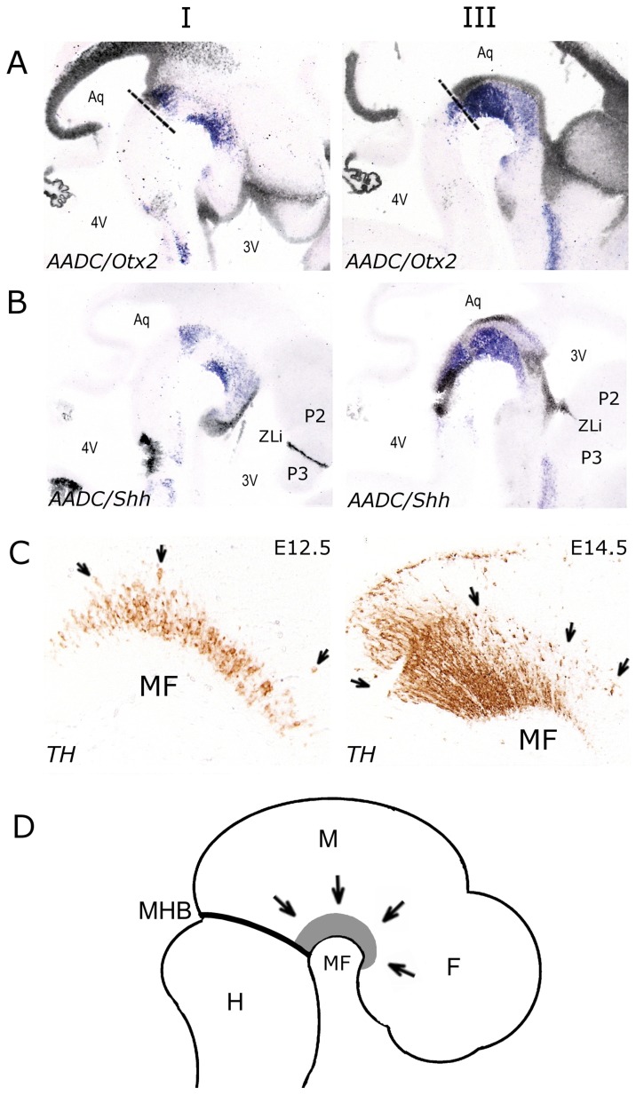
Figure 5. Analysis of ventricular zone markers Otx2 (A) and Shh (B) in E13.5 sagittal sections at two different levels, lateral (I) and medial (III).
(A) Overlays of mRNA expression patterns of Otx2 (black) and Aadc (purple) on adjacent sections. (B) Overlays of mRNA expression patterns of Shh (black) and Aadc (purple) on adjacent sections. Note that young Aadc positive neurons in the posterior diencephalon are in proximity with the ventricular zone aligning the third ventricle. (C) High magnification of the mesencephalic flexure (MF) in sagittal E12.5 and E14.5 sections immunostained with TH. Anterior is to the right and posterior to the left. Note that TH-immunoreactive neurons are radially orientated along the A/P axis (see arrows). (D) Schematic representation of a sagittal E13.5 embryo, in which mdDA neurons are depicted in gray. The arrows indicate the radial migration of DA neurons. Abbreviations: 3V, third ventricle; 4V, fourth ventricle; Aq, aqueduct; F, forebrain; H, hindbrain; M, midbrain; MF, mesencephalic flexure; MHB, mid/hindbrain border; P1, prosomere 1; P2, prosomere 2; P3, prosomere 3; ZLi, zona limitans intrathalamica.

