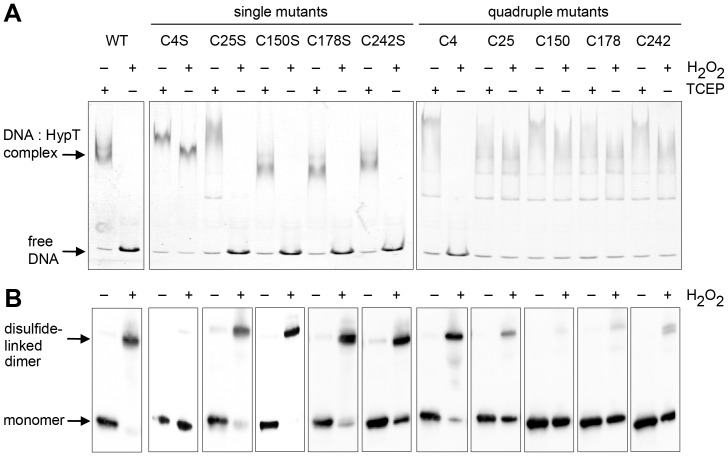
Figure 5. DNA binding and oligomerization of HypT mutants upon oxidation.
Analysis of DNA-binding activity (A) and disulfide bond formation (B) of HypT and mutants upon H2O2 treatment. HypT, single cysteine mutants, and quadruple mutants were either reduced with TCEP (1 mM) or incubated with H2O2 (1 mM) prior to incubation with DNA. A. Binding of HypT to fluorescent labeled DNA was analyzed by EMSA. Samples were separated on 6% TBE gels and DNA-protein complexes visualized using fluorescence detection. Free DNA and DNA in complex with HypT are indicated. Please note that all proteins are able to bind to DNA in the reduced state. Quadruple mutants show more diffused bands for the HypT-DNA complex, likely because they form less defined oligomers than HypT. Shown are the results of one representative experiment. B. H2O2-treated samples (without DNA) were analyzed by non-reducing SDS-PAGE. Please note disulfide-linked dimer formation at ca. 64 kDa.

