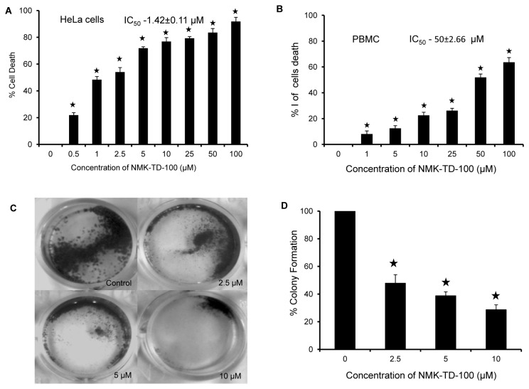
Figure 2. Cytotoxicity of NMK-TD-100 in HeLa and PBMC cells.
(A) HeLa cells were cultured for 48 h with various concentrations (0-100 µM) of NMK-TD-100. Cell viability was assessed by MTT assay and is expressed as a percentage of control. Data are represented as the mean±SD. [*p<0.05 vs. control, where n=4]. (B) Percentage of cell death in freshly isolated PBMC after treatment with NMK-TD-100 for 48 h. Data are represented as the mean±SD [*p<0.05 vs. control, where n=3]. (C, D) Colony formation assay. Cultured HeLa cells were seeded in six-well plates at a density of 1,000 cells per well and cells were treated with NMK-TD-100 (0-10 µM) for 48 h. NMK-TD-100 containing media was replaced with fresh media and subsequently cells were cultured for 10 days. At the end, cells were fixed and stained with crystal violet, images were taken. Colony formation was quantified by dissolving stained cells in Sorenson’s buffer for colorimetric reading of OD at 550 nm. Data are represented as the mean±SD [*p<0.05 vs. control, where n=3].

