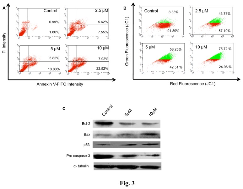
Figure 3. NMK-TD-100 induced mitochondrial pathway mediated apoptosis in HeLa cells.
(A) Annexin V-FITC /PI assay for showing that NMK-TD-100 induced apoptosis in HeLa cells. Cultured HeLa cells were treated with 0-10 µM NMK-TD-100 for 36 h, cells were harvested and stained with annexin V-FITC and PI. The percentage of early apoptotic cells in the lower right quadrant (annexin V-FITC positive/PI negative cells), as well as late apoptotic cells located in the upper right quadrant (annexin V-FITC positive/PI positive cells). (B) NMK-TD-100 induced collapse of mitochondrial membrane potential in HeLa cells. Cells were treated with 0-10 µM NMK-TD-100 for 36 h. Then treated-cells were stained with JC1 and analyzed using flow cytometer. Red fluorescence emitted from the cells with normal mitochondria (red population in figure) gradually decreases with concomitant increase in green fluorescence emitted from that containing declined mitochondrial membrane potential (green population in figure). (C) Western Blot analysis of change in expression of pro and anti-apoptotic proteins (p53, bax, bcl2 and procaspase 3) of 36 h ligand-treated HeLa cells. Probing of α- tubulin was used as a loading control. The results represent the best of data collected from three experiments with similar results.

