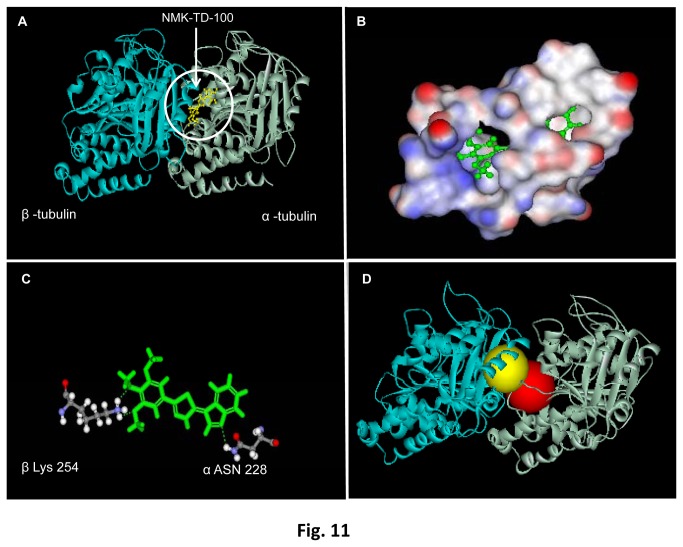
Figure 11. Docking of NMK-TD-100 to tubulin.
(A) In silico binding of NMK-TD-100 (yellow) on tubulin heterodimers. In the ribbon diagram grey represents α-tubulin monomer and blue represents β-tubulin monomer. (B) 3D diagram representing binding site of NMK-TD-100 on tubulin heterodimers. Red is showing the polar domains, blue stands for non-polar domains and white stands for neutral domains on protein surface. (C) Ball and stick figure showing positions of hydrogen bonds which stabilizes NMK-TD-100 molecule on protein surface. (D) The crystal structure of αβ heterodimer of tubulin depicting the proximity of the binding sites for colchicine (yellow) and NMK-TD-100 (red).

