. 2012 Mar;20(2):144–151. doi: 10.4062/biomolther.2012.20.2.144

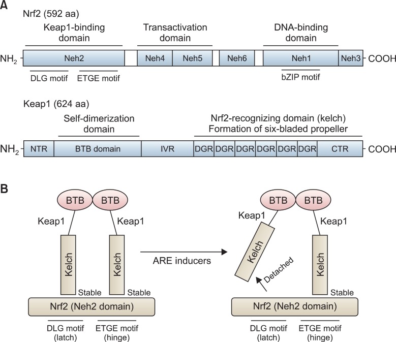
Fig. 2. Molecular architecture of Nrf2 and Keap1 proteins and two-site substrate recognition model (Hinge and Latch Model) for the Keap1/Nrf2 System. Nrf2 protein consists of 592 amino acids (592 aa) and is composed of six Neh domains (Neh1-Neh6). Neh2 is the interacting domain with Keap1 and Neh4 and Neh5 are transactivation domain. Neh1 domain contains basic leucine zipper (bZIP) motif and is the binding domain for ARE. On the other hand Keap1 consists of NTR (N-terminal Region) BTB (a Broad complex, Tramtrack and Bric a brac domain), IVR (intervening region), six DGRs (dougle glycine repeats, also called as Kelch), and CTR (carboxyl terminal region) (A). Keap1proteins homo-dimerize each other by utilizing the BTB domains in cells. The Keap1 protein homodimer recognizes the DLG (weak interaction) and ETGE (strong interaction) motifs in the Neh2 domain of Nrf2. After stress, detachment of the weak-binding DLG motif from Keap1(latch) occurs, but the strong binding ETGE motif from Keap1 (hinge), however, remains attached (B).

Fig. 2.