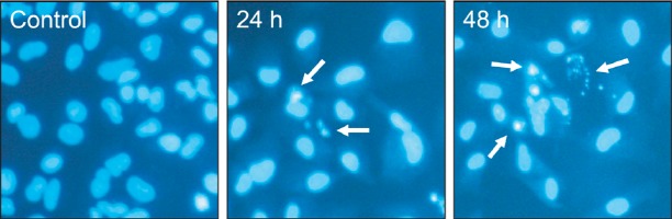
Fig. 3. TV-induced morphological changes of A549 cells. Morphological changes of A549 cell nuclei were observed by fluorescence microscopy. The cells were stained with Hoechst 33258 to identify apoptotic cells. Some of the condensed and fragmented nuclei are indicated by arrows. Data are representative of three independent experiments with similar results.