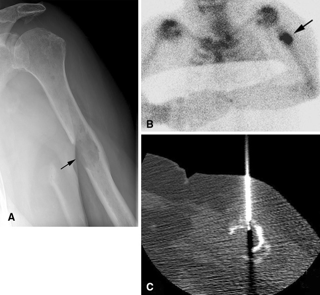
Fig. 4A–C.
A 67-year-old woman had a history of breast cancer and a painful left humerus lesion, which was nondiagnostic at core needle biopsy. Given the pain, history of malignancy, and aggressive radiographic features, surgical biopsy was performed revealing a plasmacytoma. (A) An AP radiograph shows a lytic lesion with a wide transition zone and surrounding periostitis (black arrow) in the midhumeral shaft. The (B) bone scan shows focal radiotracer uptake in the lesion (black arrow). (C) A CT-guided core needle biopsy image shows the biopsy needle in the lesion.

