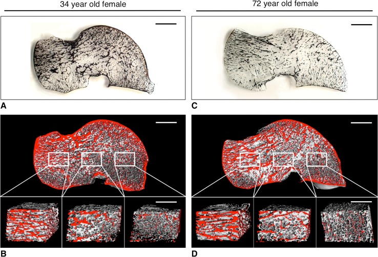
Fig. 2A–D.
Two-dimensional and three-dimensional illustrations of the age-related bone visualize the structural deterioration of the talus. The left panel indicates the two sets of plate-like trabeculae running from the trochlea tali to the (1) calcaneal facet and (2) to the neck and head of the talus in a 34-year-old woman (A, 1 mm thick, undecalcified, von Kossa-stained block grinding; bar = 1 cm). Three-dimensional visualization reveals the various structural characteristics of the respective ROIs: the body, the neck, and the talar head (B, μCT40 image, bone at the surface if colored red; bar = 1 cm). In contrast, the right panel demonstrates the predominant bone loss in the talar body of a 72-year-old woman (C, 1 mm thick, undecalcified, von Kossa stained block grinding; bar = 1 cm). However, despite age-related bone loss, three-dimensional visualization reveals the intact structural integrity of the talar neck and head (D, μCT40 image, bone at the surface if colored red; bar = 1 cm). The structural characteristics of the individual regions of interest are highlighted in the magnifications again (bar = 5 mm).

