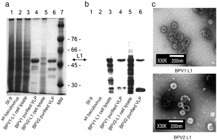
Fig. 1. Expression of major capsid protein L1 of BPV1 and BPV2 in Sf-9 insect cells.
L1 of BPV1 and BPV2 were expressed by recombinant baculoviruses and VLPs were purified by density gradient centrifugation. Crude cell lysates and purified VLPs were separated by SDS-PAGE and visualized by Coomassie staining (a), or Western blotting (b) using mAb AU-1 raised against BPV1 L1 (at a dilution of 1/10,000). Both L1 proteins migrated as 55 kD proteins. Faster migrating immunoreactive bands likely represent degradation products. Molecular weight (MW) markers are indicated. Non-infected (Sf-9) or wt baculovirus-infected cells served as controls. For transmission electron microscopy (TEM) (c) purified VLPs were negatively stained with 1% uranylacetate and visualized at a magnification × 30,000. Scale bar represents 200 nm.

