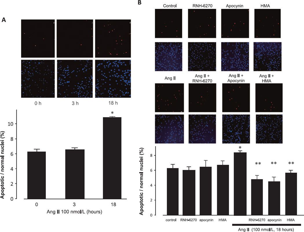
Fig. 4.
Effects of Ang II on TUNEL staining. A: Podocytes were incubated with 100 nmol/L Ang II for the indicated times. B: Podocytes were treated with 100 nmol/L Ang II for 18 h prior to TUNEL staining. Blue: DAPI stained normal nuclei and Red: TUNEL-positive nuclei. Representative photographs of TUNEL staining are shown. Data are reported as the mean ± S.E.M. (n = 6), expressed as apoptotic/normal nuclei. *P < 0.05 vs. control podocytes. **P < 0.05 vs. Ang II alone.

