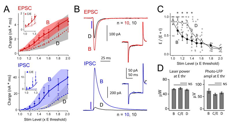
Figure 5. Effect of D-row deprivation on synaptic responses elicited by 2-ms light pulse.
A, Recruitment of recurrent EPSCs and IPSCs in spared B vs. deprived D columns. Circles, mean ± SEM. Lines and shaded region, median and 25th–75th quartiles. Insets, EPSCs and IPSCs measured in spared C and E columns (triangles), relative to B and D columns (circles and lines). B, Mean population EPSC and IPSC at 1.4 x Ethresh in B and D columns (n = 10 cells each). Inset, The average response to a −5 mV current step was not altered by deprivation. C, Fractional excitatory charge was higher in D columns, consistent with reduced IPSCs. *, p < 0.05, Tukey HSD. B column mean (not individual cells) is replotted from Fig. 3E. D, Mean effective stimulation intensity was not different between B and D columns, as assessed by laser intensity required to elicit a threshold EPSC, or by amplitude of photocurrent-LFP at Ethresh.

