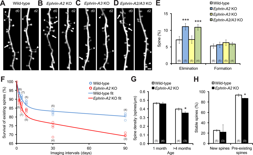
Figure 1. Dendritic Spine Elimination, but not Formation, Is Significantly Increased in One-month-old Ephrin-A2 KOs.
(A–D) Repeated imaging of the same dendritic branches over two-day intervals in the motor cortex reveals spine elimination (arrows) and formation (arrowheads), as well as filopodia (stars), in wild-type (A), ephrin-A2 KO (B), ephrin-A3 KO (C) and ephrin-A2/A3 KO (D) mice. Scale bar, 2 µm.
(E) Percentages of spines eliminated and formed over 2 days in the motor cortex of wild-type and KO mice.
(F) Percentages of spines that survived over 2, 4, 8, 30 and 90 days in wild-type and ephrin-A2 KO mice. Data were fitted by two-phase exponential decay equations.
(G) Spine density of layer V neuron apical dendrites in wild-type and ephrin-A2 KO mice in adolescence and adulthood.
(H) Survival percentages of new and pre-existing spines over 4 days in wild-type and ephrin-A2 KO mice.
Data are presented as mean ± SD. *P<0.05, ***P<0.001. See also Figures S1 and S2.

