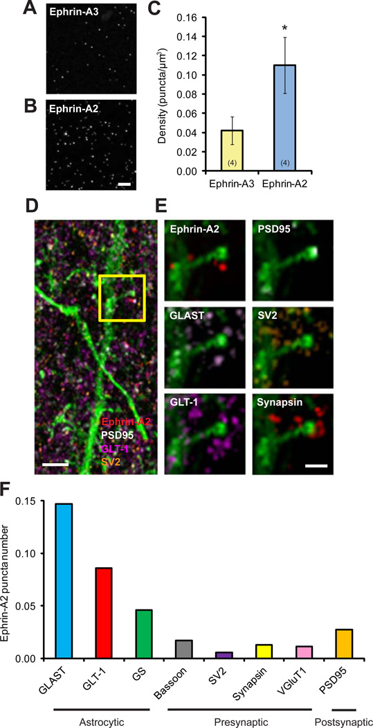
Figure 3. Ephrin-A2 Colocalizes with Astrocytic Glutamate Transporters in the Mouse Cortex.
(A, B) Representative image volumes of ephrin-A2 and ephrin-A3 immunofluorescence staining in superficial layer I of one-month-old mouse cortex. Each image is a maxium projection of 5 serial sections. Scale bar, 10 µm. Data are presented as mean ± SEM. *P<0.05.
(C) Quantification of puncta density shows that ephrin-A2 is approximately 3 times enriched, compared to ephrin-A3 in the cortex.
(D) Maximum projection of 36 serial sections of ephrin-A2 immunofluorescence (red) with protein markers of presynaptic SV2 (orange), postsynaptic PSD95 (white), astrocytic GLT-1 (magenta) and dendritic segment labeled with YFP (green). Scale bar, 1 µm.
(E) Images of a spine from the boxed region in (D) at higher magnification with various protein markers. Scale bar, 500 nm.
(F) Average numbers of ephrin-A2 puncta within 100 nm from the centers of different neuronal and astrocytic constituents.
See also Figure S4.

