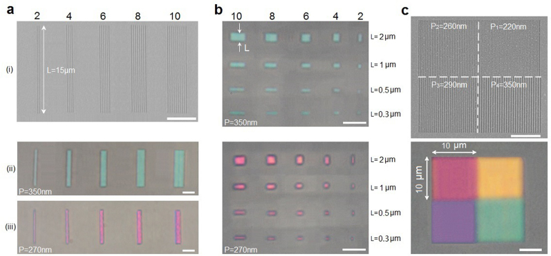
Figure 5. Ultra-compact and high-resolution plasmonic subtractive color filters.
(a) SEM image (i) of plasmonic SCFs with 2, 4, 6, 8 and 10 nanoslits of period P = 350 nm. (ii) and (iii) show the optical microscope images under TM illumination for the case of 2, 4, 6, 8 and 10 nanoslits with periods of 350 nm and 270 nm, respectively. (b) Optical microscope images of cyan (top panel, P = 350 nm) and magenta (bottom panel, P = 270 nm) plasmonic SCFs with 2, 4, 6, 8 and 10 nanoslits of differing lengths, ranging from 2 μm to 0.3 μm. (c) Top panel shows a SEM image of a plasmonic SCF mosaic consisting of four different 10 × 10 μm2 color filter squares (nanogratings with different periods of P1 = 220 nm, P2 = 260 nm, P3 = 290 nm, and P4 = 350 nm) with zero separation. Bottom panel is the corresponding optical microscope image. All of the scale bars are 5 μm.

