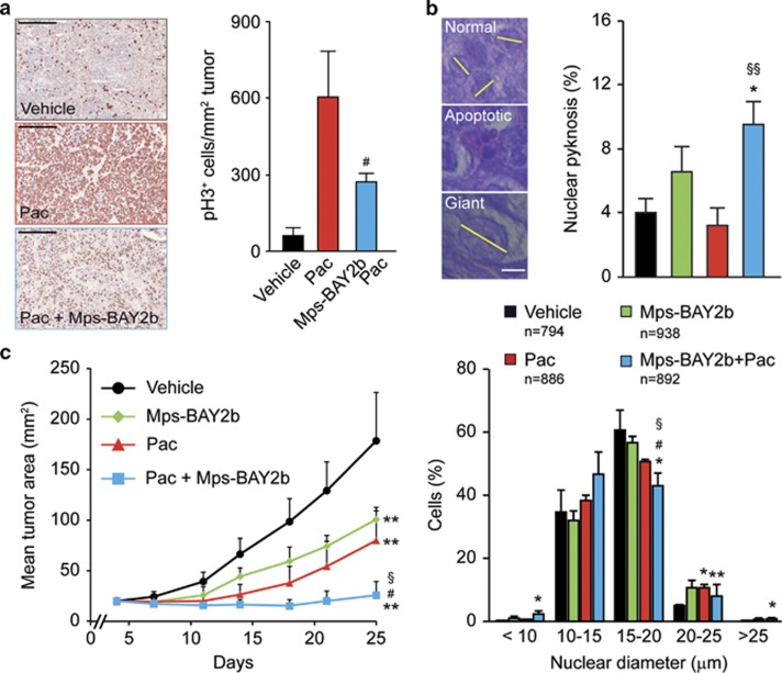
Figure 8.
Therapeutic effect of MPS1 inhibitors and paclitaxel in vivo. (a and b) Human cervical carcinoma HeLa-Matu cells were subcutaneously inoculated in athymic nu/nu mice. When tumor area reached 40–80 mm2, mice were treated with vehicle or 30 mg/kg paclitaxel (Pac) i.p., followed (after 24 h) by the administration of vehicle or the indicated dose of Mps-BAY2b p.o. (a). Alternatively, tumor-bearing mice were treated with vehicle, 8 mg/kg Pac i.v. once, 30 mg/kg Mps-BAY2b p.o. twice daily for 2 days or 8 mg/kg Pac i.v. once+30 mg/kg Mps-BAY2b p.o. twice daily for 2 days (b). (a) Tumors were recovered 1 h after the administration of Mps-BAY2b and processed for the immunohistochemical detection of phosphorylated histone 3 (pH3). Scale bar=500 μm. (b) Tumors were recovered 72 h after the first treatment and stained with hematoxylin and eosin for the light microscopy-assisted determination of nuclear diameter and nuclear pyknosis. Scale bar=10 μm. Representative microphotographs and quantitative data (means±S.E.M., n=3) are shown. (c) Athymic nu/nu mice carrying HeLa-Matu-derived xenografts were treated with vehicle, 10 mg/kg Pac i.v. once weekly, 30 mg/kg Mps-BAY2b p.o. twice daily or 10 mg/kg Pac i.v. once weekly+30 mg/kg Mps-BAY2b p.o. twice daily, and tumor area was routinely monitored by means of a common caliper. Data from one representative experiment are shown (means±S.D.). *P<0.05, **P<0.01 (Student's t-test), compared with vehicle-treated mice; #P<0.05 (Student's t-test), compared with Pac-treated mice; §P<0.05, §§P<0.01 (Student's t-test), compared with Mps-BAY2b-treated mice

