Figure 4.
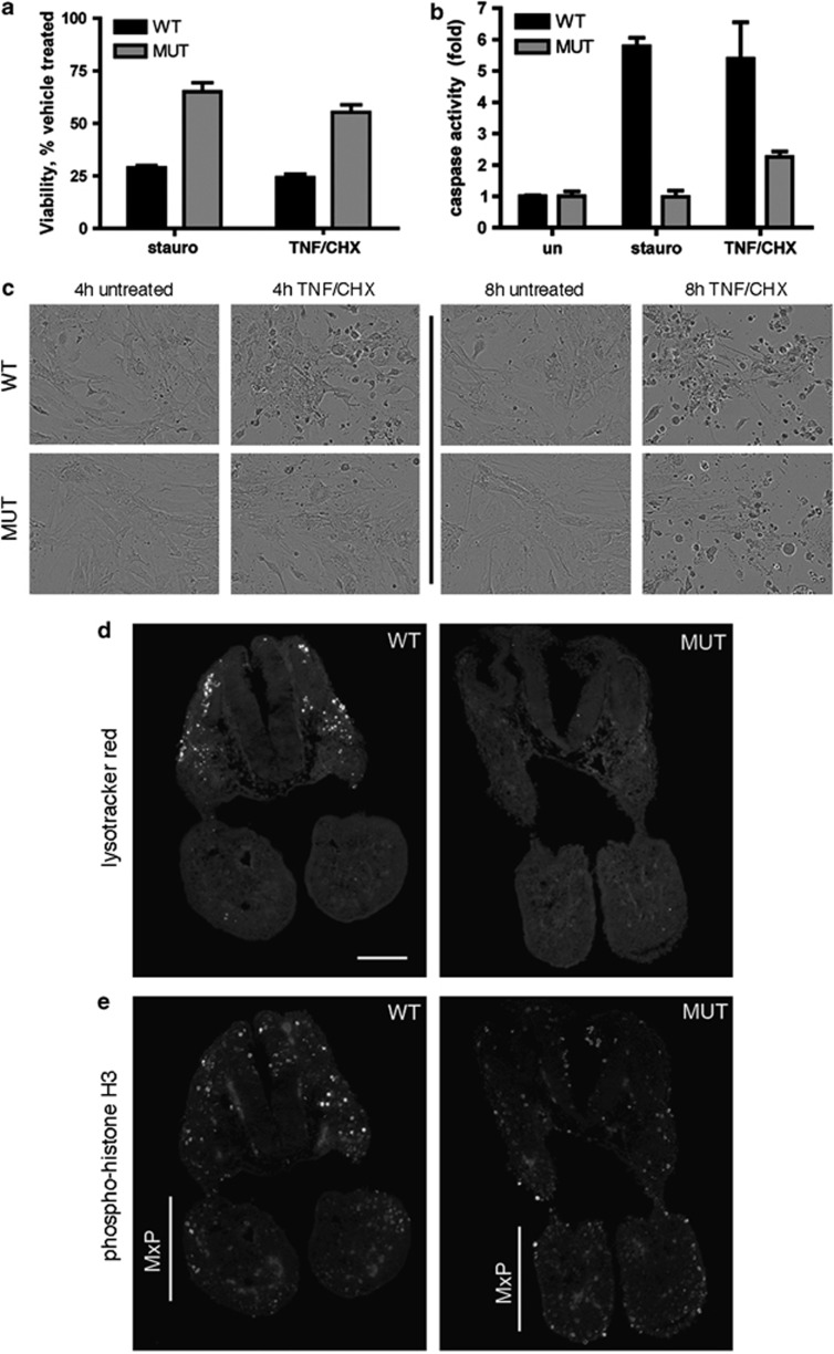
Decreased apoptosis in Apaf1yautja mutant embryos. (a) Apaf1yautja MEFs are less susceptible to death by apoptotic stimulation than wild-type cells. Viability of WT or MUT MEFs following 4-h stimulation with staurosporine (2 μg/ml) or with TNF-α (25 ng/ml) and cycloheximide (CHX; 5 μg/ml; n=4). (b) Apaf1yautja MEFs are unable to activate effector caspases. Levels of DEVDase-specific caspase activity relative to untreated WT MEFs determined 4 h after stimulation (n=3). Error bars indicate standard deviation (S.D.) of the mean. (c) Representative images of MEFs in culture 4 and 8 h after treatment with TNF and CHX. (d) LysoTracker Red staining shows decreased cell death, and (e) anti-phospho-histone H3 immunofluorescence shows increased proliferation in the maxillary prominences (MxP) of E9.5 Apaf1yautja embryos. Horizontal sections at the level of the maxillary component of the first branchial arch. Dorsal is to the top. Scale bar=100 μm

