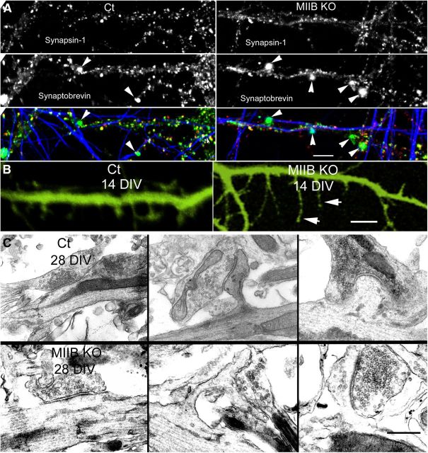
Figure 4.
MIIB KO cells show structural abnormalities at synapses. A, Comparison of immunostaining for synaptobrevin (green), synapsin-1 (red), and neurofilaments (blue) following K+ depolarization. Both Ct and MIIB KO cells show accumulations of synaptobrevin staining (arrowheads), but in the MIIB KO cells the staining frequency is increased and the accumulations are abnormally large. Scale bar, 7 μm. B, MIIB KO cells show abnormal spine development; comparison of Ct and MIIB KO cells expressing YFP. The MIIB KO cells show numerous elongated filopodia-like structures (arrowheads) and few mature spines. Phalloidin staining showed a very similar appearance. Scale bar, 2 μm. C, Comparison of synaptic development at 28 DIV. EM examples of synapses on dendritic shafts or spines. In the MIIB KO cells (bottom) the spines were abnormally long and sometimes branched. Synapses often occur on their sides. Scale bar, 350 nm.

