Figure 5.
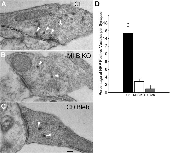
MIIB KO and blebbistatin-treated neurons show decreased percentages of HRP-positive vesicles after loading by K+ depolarization (5 min). A, EM of a thin section through a synapse from a Ct cell that was induced to take up HRP by K+ depolarization. HRP-positive vesicles (arrowheads) are numerous. B, EM of a thin section through a synapse from a MIIB KO cell that was induced to take up HRP by K+ depolarization. HRP-positive vesicles (arrowheads) are scarce. C, EM of a thin section through a synapse from a blebbistatin-treated (50 μm 30 min) Ct cell. HRP-positive vesicles (arrowheads) are rare. Scale bar, 120 nm. D, Comparison of the frequency of HRP-positive vesicles relative to the total vesicle population at each synapse. Both MIIB KO cells and blebbistatin-treated Ct cells show greatly reduced percentages of HRP-positive vesicles. The difference was significant. (ANOVA, p < 0.001; Ct, N = 14; MIIB KO, N = 14; Ct + blebbistatin, N = 12). Error bars indicate SEM.

