Figure 8.

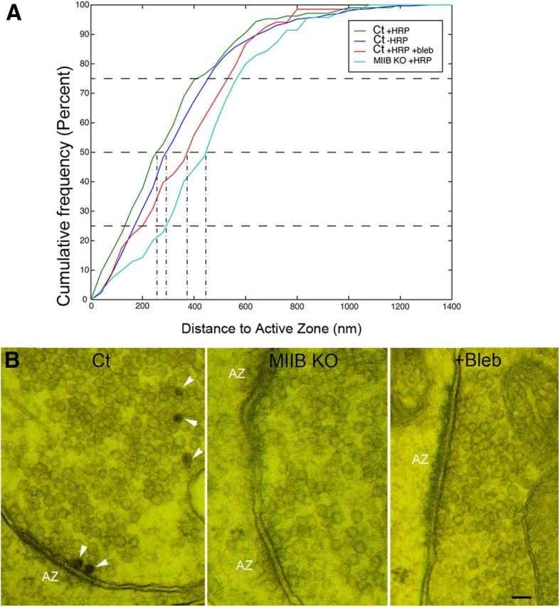
Comparison of synaptic vesicle distances to the active zone (AZ) and physical docking. A, In Ct cells the HRP-positive vesicles appear to distribute slightly closer to the AZ compared with unlabeled vesicles, but the difference was not significant (N = 14 synapses). In contrast, HRP-positive vesicles of MIIB KO and blebbistatin-treated cells appear to distribute farther from the active zone (N = 14, 12). The differences (compared with HRP-positive Ct vesicles) were significant (Kolmogorov–Smirnov test, p < 0.0001 and p = 0.02, respectively). B, Anaglyphs showing physically docked AZs. Ct cells contain HRP-positive vesicles (arrowheads). The degree of 3D depth visible in the anaglyphs varies with section thickness. Scale bar, 80 nm.
