Figure 9.
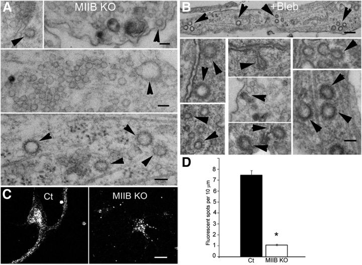
MIIB KO and blebbistatin-treated neurons show defects in CME. A, MIIB KO cells show numerous coated pits (arrowheads) following depolarization. Scale bars: 65, 65, and 130 nm, top to bottom, respectively. B, Blebbistatin treatment also causes increased numbers of coated pits. Tannic acid treatment was used to enhance coated pit staining. Scale bars: 90, 200, and 110 nm, top to bottom, respectively. C, Transferrin uptake is defective when MII activity is inhibited or decreased. Comparison of fluorescent transferrin uptake in Ct and MIIB KO hippocampal neurons. The Ct cells (left) show numerous fluorescent (Alexa 546) transferrin spots throughout the cell body and dendrites. The MIIB KO cells show sparsely distributed bright spots in the cell body. Few spots are seen in dendrites. Scale bar, 12 μm. D, Quantitation of the transferrin uptake shows the density of fluorescent transferrin spots is reduced in MIIB KO dendrites. The difference was significant (t test, N = 5 cells each from 3 cultures, p < 0.001). Error bars indicate SEM.

