
Figure 5.
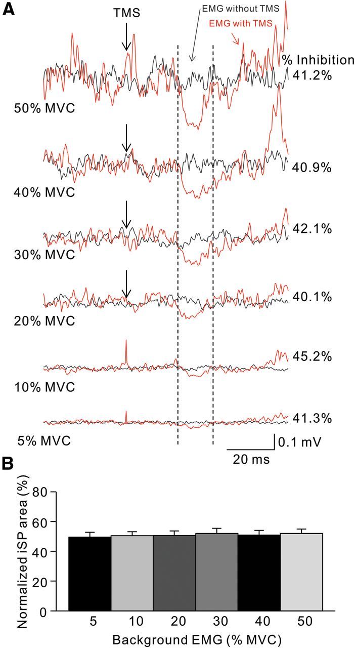
iSP during isometric index finger abduction. A, Raw traces showing rectified EMG activity in representative subjects during iSP testing at increasing levels of isometric MVC into index abduction. Traces show the average 40 trials tested with (red traces) and without (black traces) TMS. Arrows show the time of TMS during testing, and vertical dashed lines show the onset and offset of the iSP. B, Group data (n = 9). The abscissa shows all conditions tested (5, 10, 20, 30, 40, and 50% of MVC). The ordinate shows the normalized iSP area. Note that the iSP area remained unchanged during increasing levels of isometric index finger abduction. Error bars indicate SEs. *p < 0.05.