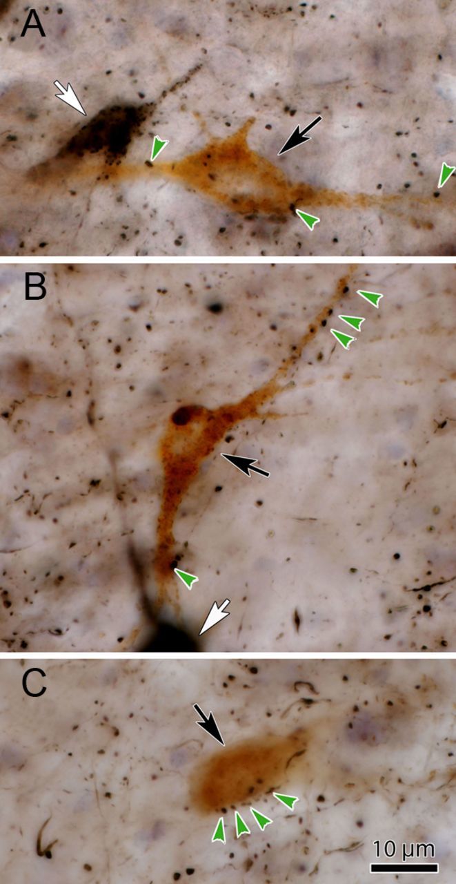
Figure 2.

Photomicrographs of the WGA-HRP-labeled neurons show close associations with BDA-labeled terminals following dual-tracer injections. A–C, The light brown WGA-HRP-labeled reticuloraphe neurons (black arrows) are easily differentiated from the black BDA-labeled reticulotectal neurons (A, B, white arrows). Close associations (green arrows) between these WGA-HRP-labeled reticuloraphe neurons and black, BDA-labeled tectoreticular boutons are common.
